New Alzheimer's Timeline
New Research Suggests Brain Changes Predate Alzheimer's Plaques by Seven Years
Anonymous
Key Points
1
Beta-amyloid is normally removed through several routes: the blood and lymphatic systems, as well as brain-resident microglia, astrocytes and enzymes. Failures across these clearance mechanisms could allow the protein to accumulate in the brain.
2
The University of Oslo study used repeated MRI scans from cognitively healthy participants collected over nearly 20 years. Researchers worked backward from the point when amyloid deposits became detectable on PET scans and compared earlier brain scans with those of participants who remained amyloid-free.
3
The Oslo findings challenge a 2024 consensus under which sufficient amyloid-beta detected in the brain is considered enough for a biological diagnosis of Alzheimer’s disease, even when a person has no symptoms.
4
Separate research links earlier menopause with faster memory decline and a greater later risk of Alzheimer’s disease; estrogen loss may be relevant because the hormone supports brain-cell function, healthy blood vessels and protection against inflammation.
5
The Oslo researchers caution that amyloid-PET, despite its sensitivity, cannot reliably detect very small or early deposits, meaning that the apparent timing of amyloid buildup may partly reflect the limits of current imaging technology.
Researchers are upending the traditional timeline of Alzheimer's disease, finding that brain changes begin years before toxic protein clumps appear Nature. A major study from the University of Oslo discovered that structural changes in the brain's outer layer were detectable at least seven years before amyloid deposits showed up on PET scans Neurology. The findings suggest Alzheimer's involves a longer chain of damage to blood vessels and waste-removal systems than previously thought.
The buildup of beta-amyloid protein happens when the brain fails to clear it through multiple routes: the bloodstream, lymphatic system, and specialized brain cells called microglia and astrocytes Nature. Failures across these clearance mechanisms may allow toxic proteins to accumulate over years, challenging the 2024 medical consensus that defines Alzheimer's by amyloid presence alone Neurology.
Structural Brain Changes Come First
The University of Oslo study tracked cognitively healthy people over nearly 20 years using repeated MRI brain scans Neurology. Researchers worked backward from when amyloid deposits became visible on PET scans and compared earlier brain images. They found that cortical thickness — the outer layer of the brain — had already changed significantly seven years before any amyloid showed up Neurology.
Rethinking How Alzheimer's Begins
The new research challenges the amyloid hypothesis, which held that toxic protein clumps spark the disease Neurology. Instead, problems in the brain's vascular system and waste-removal pathways may come first. The research suggests Alzheimer's involves a complex sequence of vascular damage, structural changes, and protein accumulation over years Nature.
The Oslo team cautions that amyloid-PET scans, while sensitive, may miss very small or early deposits Neurology. This means the apparent timing of amyloid buildup could partly reflect the limits of current imaging technology rather than the true start of the disease.
Estrogen Loss May Accelerate Decline
Separate research links earlier menopause with faster memory decline and higher Alzheimer's risk later in life Archynetys. Estrogen helps brain cells function, keeps blood vessels healthy, and protects against inflammation. When estrogen drops suddenly during early menopause, these protective effects fade faster Archynetys.
Bone Marrow's Hidden Role in Brain Health
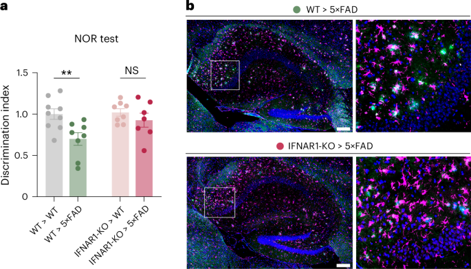
Recent findings reveal that bone marrow plays an overlooked role in Alzheimer's disease Nature. A type I interferon signal disrupts the bone marrow's production of protective immune cells called myeloid cells Nature. When this defense fails, the brain loses critical protection against inflammation and disease progression.
Sentiment
Mostly NegativeNEGATIVENEUTRALPOSITIVE
Publishers
12
Articles
2
Reach
14
A
AnonymousAuthor


